What follows is a documented clinical observation from routine radiographic evaluation at Clear Life Scoliosis and Chiropractic Center in Charlotte, NC. It is not a diagnosis. It does not establish a causal relationship. It does not conclude seizure activity or any neurological pathology. It is a pattern we are tracking in our patient population, documented here transparently, with a defined referral protocol.
If you are experiencing frequent déjà vu episodes and have not been evaluated by a neurologist, that evaluation is appropriate regardless of what you read here. Please see your primary care physician or a neurologist directly. This page is not a substitute for that evaluation.
Clinical Observation · Upper Cervical · Charlotte, NC
Atlas Position and Déjà Vu Frequency — A Clinical Observation From Clear Life Scoliosis Charlotte NC
Clinical observations do not always arrive neatly packaged. Sometimes they emerge from a pattern that becomes too consistent to ignore and not yet sufficiently documented to publish. This is one of those observations. We are documenting it publicly because we believe clinicians and patients deserve to know what we are seeing — with complete transparency about what it is and what it is not.
At Clear Life Scoliosis and Chiropractic Center in Charlotte, NC, every scoliosis and spinal rehabilitation patient undergoes quantitative lateral cervical radiographic analysis as part of the standard evaluation. That analysis includes measurement of the atlas — C1 — angle relative to the horizontal plane. In the course of systematic intake assessment, a pattern has emerged that we are actively tracking and intend to pursue through formal research publication.
Patients whose atlas angle measures below 18 degrees on lateral cervical radiograph — indicating a more horizontal atlas position — are reporting multiple déjà vu episodes per month at a frequency that appears disproportionate to what we observe in patients whose atlas angle is within a more typical range. The pattern is consistent enough across our patient population that it warrants documentation and referral protocol. It is not consistent enough — and not yet formally studied — to support a clinical conclusion.
What We Are Measuring — The Atlas Angle on Lateral Cervical Radiograph
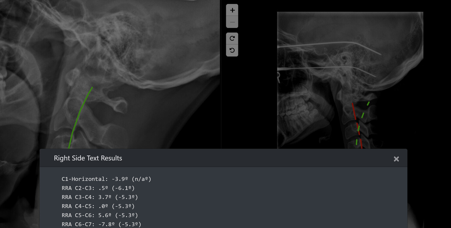
The atlas — C1 — is the first cervical vertebra. It sits directly beneath the occiput and articulates with the base of the skull at the atlanto-occipital joint. Its position relative to the horizontal plane is measurable on a standing lateral cervical radiograph using the inferior endplate of the atlas as the reference line.
In standard cervical radiographic analysis, the atlas angle — the inclination of the C1 inferior endplate relative to the horizontal — varies across individuals. A more lordotic upper cervical curve produces a more inclined atlas. A flatter or kyphotic upper cervical segment produces a more horizontal atlas position.
The threshold we are tracking is 18 degrees. Patients whose atlas inferior endplate angle falls below 18 degrees from horizontal on standing lateral cervical radiograph represent a more horizontal atlas position. At Clear Life Scoliosis Charlotte NC, this measurement is performed using PostureRay software, which carries published inter- and intra-rater reliability data. The measurement is quantitative, reproducible, and taken in the standing weight-bearing position — not supine. Dr. Justin M. Dick, DC holds CNMT and ARRT(N)(CT) dual imaging credentials that inform how these measurements are performed and interpreted. See Radiographic Imaging Credentials Charlotte NC.
This measurement is also directly relevant to the published research from Clear Life Scoliosis — Dick JM, Cureus 2025, PMID 41018459 — which found abnormal C3-C4 cervical segmental motion in over 70% of structural spinal injury patients on stress radiography. The atlas position is evaluated as part of the same systematic upper cervical radiographic analysis in which that finding was identified. The clinical picture of upper cervical mechanical dysfunction in scoliosis patients is broader than a single measurement — the atlas angle below 18 degrees is one data point within that picture.
What We Are Observing — The Pattern
Patients presenting to Clear Life Scoliosis and Chiropractic Center in Charlotte, NC with a lateral cervical radiograph demonstrating atlas angle below 18 degrees from horizontal are reporting multiple déjà vu episodes per month on structured intake assessment at a frequency that appears disproportionate to patients whose atlas angle measures above 18 degrees.
This is an observed pattern in a clinical population. It has not been formally studied. It has not been controlled for confounding variables. It does not establish causation. It does not permit a clinical diagnosis of any condition. It is being documented here as a clinical observation and as the basis for a defined referral protocol.
Déjà vu — the subjective experience of familiarity with a present situation that objectively could not have been encountered before — is a common transient phenomenon in the general population. Occasional déjà vu is not clinically significant in isolation. Frequent, recurring déjà vu — multiple episodes per month — is a different clinical picture. Frequent déjà vu is a documented feature of temporal lobe epilepsy and other neurological conditions. It is also reported in migraine, anxiety disorders, sleep deprivation, and as a side effect of various medications. It can occur without any identifiable pathology.
We are not concluding that the atlas position is causing déjà vu. We are not concluding that our patients have temporal lobe epilepsy or any seizure disorder. We are observing that within our patient population — which is enriched for structural spinal pathology and upper cervical mechanical dysfunction relative to the general population — patients with this specific radiographic finding are reporting this specific symptom at a frequency that has become clinically noticeable. That observation is the entirety of the claim.
Why the Atlas Position May Be Relevant — The Mechanistic Hypothesis
This section describes a biological hypothesis — not a proven mechanism. It is offered to provide clinical context for why this observation might be worth pursuing formally, not to establish causation.
The atlas articulates with the occiput at the atlanto-occipital joint and with C2 at the atlanto-axial joint. The upper cervical spine — C0 through C2 — has a unique relationship with the brainstem, the vertebral arteries, and the upper cervical proprioceptive system that is not shared by the lower cervical levels. The vertebral arteries transit through the transverse foramina of C1 and C2 before entering the foramen magnum. The atlanto-occipital and atlanto-axial joints contain the highest density of mechanoreceptors in the cervical spine, providing substantial proprioceptive input to the cerebellum and brainstem.
A horizontally positioned atlas — flatter than typical relative to the skull base — alters the biomechanical relationship between C1, C2, and the occiput. Whether and how that altered relationship might influence vertebral artery hemodynamics, upper cervical proprioceptive afferent signaling, or brainstem function is not established. There is published literature on the relationship between upper cervical dysfunction and headache, dizziness, and tinnitus — conditions with some mechanistic overlap with temporal lobe function — but that literature does not directly address the atlas angle measurement or the déjà vu symptom specifically.
The hypothesis is mechanistically plausible. It is not proven. The observation that prompted it requires formal study before any clinical conclusion is appropriate.
What We Do With This Finding — The Referral Protocol
When a patient at Clear Life Scoliosis and Chiropractic Center presents with an atlas angle below 18 degrees on lateral cervical radiograph and reports multiple déjà vu episodes per month on structured intake assessment, the following steps are taken:
- The radiographic finding is documented with the quantitative measurement and the PostureRay measurement methodology in the patient record.
- The symptom frequency is documented on the intake assessment with the specific number of episodes per month reported.
- The patient is informed that the combination of this radiographic finding and symptom frequency warrants neurological evaluation — not because a diagnosis has been made but because the pattern is one a neurologist should assess.
- A written referral recommendation to a neurologist or the patient's primary care physician is provided.
- The patient's structural spinal rehabilitation continues as clinically indicated — the referral does not interrupt or replace chiropractic care for the spinal condition that brought the patient to Clear Life.
This referral protocol operates entirely within chiropractic scope of practice. Identifying a radiographic finding, documenting an associated symptom pattern, and recommending appropriate specialist referral is a standard component of clinical practice. No diagnosis is being made. No neurological treatment is being provided or recommended at Clear Life.
What Déjà Vu Actually Is — Clinical Background
Déjà vu is a French phrase meaning "already seen." It describes the experience of perceiving a current situation as having been encountered before, despite knowing objectively that it could not have been. The experience is typically brief — seconds to a minute — and is accompanied by a strong sense of familiarity without the ability to identify what specifically feels familiar.
Déjà vu as an isolated, infrequent experience is common. Surveys of the general population find that the majority of people have experienced déjà vu at least once. The clinical significance of déjà vu depends on its frequency, duration, associated features, and clinical context.
Frequent déjà vu — multiple episodes per month — appearing as part of a seizure aura is a recognized feature of temporal lobe epilepsy, specifically simple partial seizures arising from the hippocampus or parahippocampal gyrus. These episodes may occur with or without subsequent convulsion. A patient experiencing frequent déjà vu — particularly if episodes are stereotyped, associated with other perceptual changes, or followed by confusion — warrants neurological evaluation including EEG.
Frequent déjà vu also occurs in the context of migraine with aura, anxiety disorders, depersonalization-derealization disorder, sleep deprivation, and medication side effects. The neurological workup is designed to distinguish between these possibilities — not to assume epilepsy as the default explanation.
We are not characterizing our patients' déjà vu experiences as seizure auras. We are observing that frequent déjà vu in the context of a specific radiographic finding in a structured spinal rehabilitation population is a pattern that warrants a neurologist's evaluation.
The Scoliosis Context — Why This Population
The patient population at Clear Life Scoliosis and Chiropractic Center is not the general population. Our patients are evaluated specifically for structural spinal pathology — scoliosis, post-collision cervical injury, and complex spinal presentations. Every patient receives systematic upper cervical radiographic analysis as part of the standard evaluation. That analysis is what made this pattern visible.
In the general chiropractic practice, atlas angle is not routinely measured quantitatively. Patients are not routinely asked about déjà vu frequency on structured intake. The combination of systematic upper cervical radiographic measurement and structured symptom assessment is what created the clinical conditions in which this pattern became observable. Without both components, the association would remain invisible.
Published research from Clear Life Scoliosis — PMID 41018459, the cervical mechanics paper — found that upper cervical mechanical dysfunction is far more prevalent in the scoliosis population than standard clinical evaluation identifies. That finding is the research foundation for why upper cervical systematic assessment is standard at every evaluation here. The atlas angle observation emerged from that same systematic assessment framework.
The scoliosis population may have specific reasons to develop upper cervical mechanical dysfunction — the rotational and sagittal plane changes of scoliosis produce compensatory adaptations in the upper cervical spine that are well-documented in the structural rehabilitation literature. Whether those compensatory adaptations produce atlas angle changes that are specifically associated with neurological symptom frequency is the question that formal research would need to address.
The Path Forward — Toward Formal Publication
A clinical observation documented on a blog post is not evidence. It is the preliminary step before evidence — the documented observation that justifies designing a formal study. We are being explicit about that distinction because it matters.
The intention is to pursue formal peer-reviewed publication of this observation. The appropriate study design is a retrospective chart review of patients seen at Clear Life Scoliosis Charlotte NC with quantitative atlas angle measurement available — identifying the proportion reporting frequent déjà vu in the below-18-degree group versus the above-18-degree group, controlling for the confounding variables that independently predict déjà vu frequency, and reporting the finding with appropriate statistical analysis and honest discussion of limitations.
That study, if it confirms the pattern, would be the first published data on this specific association. If it does not confirm the pattern at a statistically significant level, that result is also publishable and equally important. The commitment is to formal study and honest reporting — not to confirmation of the observation.
Dr. Justin M. Dick, DC has multiple PubMed-indexed publications in Cureus. The research infrastructure for this study is in place. The timeline for submission depends on the chart review process and institutional considerations. Colleagues who have seen similar patterns in their own upper cervical or scoliosis patient populations are encouraged to reach out through the contact information below.
Clinician outreach.
If you are a clinician who performs routine quantitative upper cervical radiographic analysis and has observed frequent déjà vu reporting in patients with specific atlas position measurements, Dr. Justin M. Dick, DC at Clear Life Scoliosis and Chiropractic Center in Charlotte, NC welcomes collegial discussion of this observation. Contact: office@clearlifescoliosis.com
If You Are a Patient Reading This
If you found this page because you are experiencing frequent déjà vu and are trying to understand what it means, there are a few things worth being clear about.
Frequent déjà vu warrants medical evaluation. That evaluation should begin with your primary care physician, who can assess whether a neurology referral is appropriate. If you have a known scoliosis diagnosis and have not had a systematic upper cervical radiographic evaluation, that evaluation at Clear Life Scoliosis Charlotte NC would document your atlas angle and add a specific radiographic data point to the clinical picture you bring to your neurologist.
This page does not diagnose you. It does not predict your neurological status. It does not replace a physician or neurologist evaluation. It documents a clinical pattern we are tracking and a referral protocol we follow when we identify it. If you are concerned about your déjà vu frequency, the right response is to contact your doctor — not to rely on this observation as an explanation.
If you have a scoliosis diagnosis and would like a systematic upper cervical radiographic evaluation at Clear Life Scoliosis Charlotte NC — 8814 Rachel Freeman Way Suite 103, Charlotte NC 28278 — Dr. Justin M. Dick, DC holds CNMT and ARRT(N)(CT) dual imaging credentials and performs quantitative atlas angle measurement as part of the standard scoliosis evaluation. No referral required. Call 980-368-0766.
Quantitative Upper Cervical Radiographic Evaluation at Clear Life Charlotte
Dr. Justin M. Dick, DC — CNMT · ARRT(N)(CT) · CLEAR Fellow · CLEAR Board of Directors · Multiple PubMed-indexed publications on cervical mechanics in scoliosis patients. Quantitative atlas angle measurement using PostureRay as part of every scoliosis evaluation. No referral required. Cash-based practice.
Charlotte, NC 28278
980-368-0766 | office@clearlifescoliosis.com
Book Online at JaneApp
Justin Dick
Contact Me