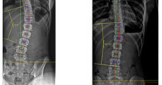
A healthy spine is straight from the front and curved from the side. Seen from the side, the neck and low back curve inward (lordosis) and the mid back curves outward (kyphosis). Seen from the front, any sideways curve of 10° or more measured by the Cobb method is scoliosis. The side-view curves have no single normal number. Bernhardt and Bridwell measured 102 radiographically normal spines and found a wide range of normal sagittal alignment, and Been and Kalichman, after reviewing more than 120 papers, concluded that normal lordosis depends on age, sex, body mass, ethnicity, and sport.
I am Dr. Justin Dick at Clear Life Scoliosis and Chiropractic Center in Charlotte, NC. I hold CNMT and ARRT(N)(CT) imaging credentials, and reading side-view alignment is part of every spinal evaluation in our office. This post explains what normal looks like and where the evidence on "abnormal" is strong and where it is thin.
The Three Side-View Curves
Cervical lordosis is the inward curve of the neck. Thoracic kyphosis is the outward curve of the rib cage region. Lumbar lordosis is the inward curve of the low back. Bernhardt and Bridwell found the thoracolumbar junction, where the rib cage meets the low back, is close to straight in normal spines, and that lumbar lordosis usually starts at L1-L2 and increases at each level down to the sacrum. Most of the low back's curve sits in its lowest two or three segments.
Kyphosis vs Lordosis
Both words describe direction, not disease. Kyphosis is a curve that bows backward and lordosis is a curve that bows forward. The terms become diagnoses only when the curve is outside the expected range for that person or sits in the wrong region. A lordotic thoracic spine or a kyphotic lumbar spine is abnormal by location regardless of the angle. Clément and colleagues studied 86 patients with thoracic adolescent idiopathic scoliosis and found that reduced thoracic kyphosis (hypokyphosis) was independent of pelvic shape and behaved as a structural part of the scoliotic deformity. A flat mid back on a side-view film in a teenager is one reason I look harder at the front view.
Why There Is No Single Normal Number
Fon and colleagues measured thoracic kyphosis on 316 normal subjects aged 2 to 77 and found it increased with age, faster in women than men. A kyphosis angle that is normal at 70 can be excessive at 15. Roussouly and colleagues studied 160 volunteers without spinal symptoms and showed that the shape of the lumbar lordosis follows the tilt of the top of the sacrum, which is set by pelvic anatomy. Two people with healthy spines can have very different lordosis angles because their pelvises differ. A report that says "loss of lordosis" with no reference to pelvic parameters is a description, not a diagnosis.
When Side-View Alignment Matters Clinically
The strongest evidence concerns overall balance in adults. Glassman and colleagues analyzed 752 adults with spinal deformity, 352 with positive sagittal balance, meaning the head sits forward of the pelvis on a standing film. Every health status measure worsened as the forward shift increased, and kyphosis in the lumbar region was poorly tolerated. Chun and colleagues pooled 13 studies with 796 patients with low back pain and 927 controls and found smaller lumbar lordosis in the pain group, especially in disc herniation and degeneration. Those were observational studies. They show association, and a painful disc can flatten the low back as easily as a flat back can load the disc.
What a Radiograph Can and Cannot Tell You
A standing full-spine radiograph shows the curves under load. A lying MRI does not show standing alignment, so it cannot answer a sagittal balance question. Measurement method matters too: Been and Kalichman noted that arm position and the vertebrae included change the lordosis angle, which is one reason two reports on the same spine can disagree. My post on scoliosis misdiagnosis covers measurement error.
How Dr. Justin Dick Evaluates Spinal Curves at Clear Life
At our Charlotte office, the evaluation includes standing front and side radiographs when clinically indicated, Cobb angle measurement in the front view, regional side-view angles, and global balance, read against the patient's age and pelvic shape. Our page on cervical alignment in scoliosis covers the neck, which is often overlooked.
Comparison
| Region | Normal direction (side view) | What the evidence says | Source |
|---|---|---|---|
| Neck | Lordosis (inward) | Abnormal cervical mechanics analyzed in scoliosis patients | Dick 2025, Cureus |
| Mid back | Kyphosis (outward) | Increases with age, faster in women | Fon 1980, 316 subjects |
| Thoracolumbar junction | Close to straight | Lordosis starts at L1-L2 | Bernhardt and Bridwell 1989, 102 spines |
| Low back | Lordosis (inward) | Shape follows sacral slope and pelvic anatomy | Roussouly 2005, 160 volunteers |
| Global balance | Head over pelvis | Health scores worsen as forward shift increases | Glassman 2005, 752 adults |
Research and Evidence
Evidence tiering: Bernhardt, Fon, and Roussouly are cross-sectional radiographic studies of normal subjects. Glassman is a retrospective analysis of a prospective deformity database. Chun is a meta-analysis of observational studies and cannot establish cause. Been and Kalichman is a narrative review. No source here gives a single normal angle that applies to every patient, and I do not quote one.
My own published work addresses side-view alignment directly. The cervical mechanics study, A Retrospective Cross-Sectional Analysis of Abnormal Cervical Mechanics in Patients With Scoliosis (Cureus, 2025, PMID 41018459), analyzed neck alignment in scoliosis patients, and the geriatric sagittal alignment case series, Radiographic Sagittal Alignment and Kinetic Chain Alterations in Geriatric Patients With Scoliosis: A Case Series (Cureus, 2026, PMID 42037873), describes side-view alignment in two older adults. Both are observational and case-level evidence.
References
- Bernhardt M, Bridwell KH. Segmental analysis of the sagittal plane alignment of the normal thoracic and lumbar spines and thoracolumbar junction. Spine. 1989;14(7):717-21. doi:10.1097/00007632-198907000-00012
- Fon GT, Pitt MJ, Thies AC. Thoracic kyphosis: range in normal subjects. AJR Am J Roentgenol. 1980;134(5):979-83. doi:10.2214/ajr.134.5.979
- Been E, Kalichman L. Lumbar lordosis. Spine J. 2014;14(1):87-97. doi:10.1016/j.spinee.2013.07.464
- Roussouly P, Gollogly S, Berthonnaud E, Dimnet J. Classification of the normal variation in the sagittal alignment of the human lumbar spine and pelvis in the standing position. Spine. 2005;30(3):346-53. doi:10.1097/01.brs.0000152379.54463.65
- Glassman SD, Bridwell K, Dimar JR, et al. The impact of positive sagittal balance in adult spinal deformity. Spine. 2005;30(18):2024-9. doi:10.1097/01.brs.0000179086.30449.96
- Chun SW, Lim CY, Kim K, Hwang J, Chung SG. The relationships between low back pain and lumbar lordosis: a systematic review and meta-analysis. Spine J. 2017;17(8):1180-1191. doi:10.1016/j.spinee.2017.04.034
- Clément JL, Geoffray A, Yagoubi F, et al. Relationship between thoracic hypokyphosis, lumbar lordosis and sagittal pelvic parameters in adolescent idiopathic scoliosis. Eur Spine J. 2013;22(11):2414-20. doi:10.1007/s00586-013-2852-z
- Dick JM. A retrospective cross-sectional analysis of abnormal cervical mechanics in patients with scoliosis. Cureus. 2025;17(8):e91098. doi:10.7759/cureus.91098
- Whelan JP, Dick JM. Radiographic sagittal alignment and kinetic chain alterations in geriatric patients with scoliosis: a case series. Cureus. 2026;18(3):e105827. doi:10.7759/cureus.105827
Frequently Asked Questions
What is a normal curvature of the spine?
From the front, the spine should be straight. From the side, the neck and low back curve inward (lordosis) and the mid back curves outward (kyphosis). Published studies of normal spines show a wide range of normal side-view angles.
What is the difference between kyphosis and lordosis?
Kyphosis is a curve that bows backward, normal in the mid back. Lordosis is a curve that bows forward, normal in the neck and low back. Either becomes a problem when it is outside the expected range or in the wrong region.
Is there one normal degree of lordosis?
No. A review of more than 120 papers found that normal lordosis varies with age, sex, body mass, ethnicity, and sport, and that lordosis shape follows pelvic anatomy.
Does losing the curve in my low back cause pain?
A 2017 meta-analysis found people with low back pain had smaller lumbar lordosis than controls, especially with disc problems. The studies were observational, so they cannot show which came first.
Can an MRI show my spinal curves?
Not reliably. MRI is taken lying down. Standing radiographs show the curves under load, which is what alignment assessment needs. Dr. Justin Dick at Clear Life Scoliosis in Charlotte, NC reads both.
Where can I get a spinal alignment evaluation in Charlotte, NC?
Clear Life Scoliosis and Chiropractic Center, 8814 Rachel Freeman Way, Suite 103, Charlotte, NC 28278, provides radiographic alignment evaluation with Dr. Justin Dick, DC, CNMT, ARRT(N)(CT). Call 980-368-0766.
Service Area
Clear Life Scoliosis and Chiropractic Center sees patients from Charlotte NC, Huntersville NC, Ballantyne NC, Matthews NC, Concord NC, Mooresville NC, Rock Hill SC, Fort Mill SC. Families from across the Charlotte region and the South Carolina border communities come to the Rachel Freeman Way office for radiographic scoliosis evaluation with Dr. Justin Dick.
Related Pages
- Scoliosis Biomechanics and Sagittal Alignment
- Cervical Alignment in Scoliosis
- Kyphosis Treatment in Charlotte
- Hyperkyphosis Treatment
- Scoliosis Treatment for Adults
- Postural vs Structural Scoliosis
- Cobb Angle Measurement
- Tech Neck
- Scoliosis Imaging and Measurement
- Dr. Justin Dick Research
Schedule an Evaluation in Charlotte
If a report mentions loss of lordosis, kyphosis, or imbalance and you want it explained against your own anatomy, Dr. Justin Dick reviews standing films at our Charlotte office.
Dr. Justin M. Dick, DC
8814 Rachel Freeman Way, Suite 103, Charlotte, NC 28278
Phone: 980-368-0766 · Email: office@clearlifescoliosis.com
clearlifescoliosis.com · Book online
Justin Dick
Contact Me