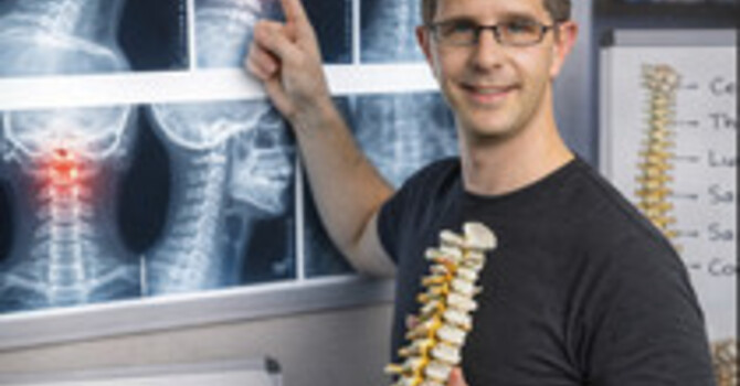
A standing full-spine radiograph is the standard way to diagnose and measure scoliosis, because it shows the Cobb angle under load. Radiation is a real consideration in growing patients who need repeat films. In the US Scoliosis Cohort, 5,573 women diagnosed before age 20 between 1912 and 1965 had an average of 24.7 radiographs, and breast cancer deaths were 69 percent higher than expected, rising with dose. In a Danish cohort of 215 patients treated between 1983 and 1990 with an average of 16 radiographs, cancer risk was 4.8 times that of the general population. Modern low-dose systems reduce dose substantially: EOS slot-scanning cut skin dose 6 to 9 times in one comparison.
I am Dr. Justin M. Dick, DC, at Clear Life Scoliosis and Chiropractic Center in Charlotte, NC. I hold CNMT and ARRT(N)(CT) credentials in nuclear medicine and CT, so dose and image quality are part of my training. This post explains both sides.
Which Views Are Needed
A standing posteroanterior (back-to-front) full-spine view measures the Cobb angle and shows the pelvis for growth staging. A lateral view shows sagittal alignment. Bending views help judge flexibility before surgery or bracing decisions. Not every visit needs every view. My post on Cobb angle measurement explains what is measured.
What the Long-Term Studies Found
Doody and colleagues followed 5,466 women from the US Scoliosis Cohort for an average of 40 years. Mean estimated breast dose was 10.8 cGy from older techniques. There were 77 breast cancer deaths against 45.6 expected (standardized mortality ratio 1.69), and risk rose with number of exposures and cumulative dose. The authors noted that curve severity and reproductive history could confound part of the finding. Simony and colleagues followed 215 Danish patients treated from 1983 to 1990. Nine developed cancer, mostly breast and endometrial, a relative risk of 4.8; they estimated 0.8 to 1.4 mSv per examination, which they described as comparable to modern equipment. That is a small cohort with wide uncertainty.
How Dose Is Reduced
Deschênes and colleagues compared EOS slot-scanning imaging with conventional computed radiography in 50 scoliosis patients. Skin dose in the thoracoabdominal region was 6 to 9 times lower with EOS, and image quality was better for most structures. Other practical measures: posteroanterior positioning instead of anteroposterior reduces dose to the breasts, collimation limits the exposed area, shielding where it does not obscure anatomy, and spacing films by clinical need. Those practices are standard radiation protection principles.
Can a Scoliometer Replace X-Rays?
For screening between films, a scoliometer or surface measurement helps. It cannot replace a radiograph for measuring the curve; my scoliometer post covers its accuracy limits.
How Dr. Justin M. Dick, DC Handles Imaging at Clear Life
At our Charlotte office, radiographs are ordered when they will change a decision, using posteroanterior positioning and collimation, and spaced to the growth stage and treatment phase. Prior films are requested to avoid repeats. Our imaging credentials page and imaging and measurement page describe the approach.
Comparison
| Study | Population | Finding |
|---|---|---|
| Doody 2000 | 5,466 women, diagnosed 1912 to 1965 | Breast cancer mortality ratio 1.69; risk rose with dose |
| Simony 2016 | 215 patients, treated 1983 to 1990 | Cancer relative risk 4.8; mean 16 radiographs |
| Deschênes 2010 | 50 patients, EOS vs CR | Skin dose 6 to 9 times lower with EOS |
Research and Evidence
Evidence tiering: Doody and Simony are retrospective cohorts; both may be confounded by disease severity and other factors, and Simony is small. Deschênes is a comparative imaging study. Dose-reduction practices listed are standard radiation protection principles.
My own published work relies on standing radiographic measurement. The 13-month follow-up case, Reduction of Adolescent Idiopathic Scoliosis: A 13-Month Follow-Up (Cureus, 2025, PMID 40062184), reports radiographic Cobb angle change in one adolescent.
References
- Doody MM, Lonstein JE, Stovall M, et al. Breast cancer mortality after diagnostic radiography: findings from the U.S. Scoliosis Cohort Study. Spine. 2000;25(16):2052-63. doi:10.1097/00007632-200008150-00009
- Simony A, Hansen EJ, Christensen SB, Carreon LY, Andersen MO. Incidence of cancer in adolescent idiopathic scoliosis patients treated 25 years previously. Eur Spine J. 2016;25(10):3366-3370. doi:10.1007/s00586-016-4747-2
- Deschênes S, Charron G, Beaudoin G, et al. Diagnostic imaging of spinal deformities: reducing patients radiation dose with a new slot-scanning X-ray imager. Spine. 2010;35(9):989-94. doi:10.1097/BRS.0b013e3181bdcaa4
- Dick JM, Spurgeon S. Reduction of adolescent idiopathic scoliosis: a 13-month follow-up. Cureus. 2025;17(2):e78669. doi:10.7759/cureus.78669
Frequently Asked Questions
Are scoliosis X-rays dangerous?
Repeated radiographs in childhood carry some cancer risk. A US cohort of women diagnosed between 1912 and 1965 had 69 percent higher breast cancer mortality than expected, rising with dose.
How many X-rays does scoliosis need?
It depends on growth and treatment phase. Dr. Justin M. Dick, DC at Clear Life Scoliosis in Charlotte, NC orders films only when they will change a decision and requests prior films to avoid repeats.
What is EOS imaging?
A low-dose slot-scanning system. In one comparison, skin dose was 6 to 9 times lower than conventional radiography with better image quality for most structures.
Why is a back-to-front X-ray used for scoliosis?
Posteroanterior positioning reduces dose to the breasts compared with front-to-back positioning, a standard radiation protection practice.
Can a scoliometer replace an X-ray?
No. It is useful for screening between films but cannot measure the Cobb angle.
Where can I get scoliosis X-rays reviewed in Charlotte, NC?
Clear Life Scoliosis and Chiropractic Center, 8814 Rachel Freeman Way, Suite 103, Charlotte, NC 28278, provides radiographic review with Dr. Justin M. Dick, DC, CNMT, ARRT(N)(CT). Call 980-368-0766.
Service Area
Clear Life Scoliosis and Chiropractic Center sees patients from Charlotte NC, Huntersville NC, Ballantyne NC, Matthews NC, Concord NC, Mooresville NC, Rock Hill SC, Fort Mill SC. Families from across the Charlotte region and the South Carolina border communities come to the Rachel Freeman Way office for radiographic scoliosis evaluation with Dr. Justin M. Dick, DC.
Related Pages
- Radiographic Imaging Credentials
- Scoliosis Imaging and Measurement
- Cobb Angle Measurement
- Can Scoliosis Be Misdiagnosed
- Spinal Rotation in Scoliosis
- Carolinas Scoliosis Screening Initiative
- Free Scoliosis Screening Tool
- When Scoliosis Progresses the Most
- Scoliosis Care Program
- Dr. Justin M. Dick, DC Research
Schedule an Evaluation in Charlotte
If you are worried about how many films your child has had, bring the history and Dr. Justin M. Dick, DC will plan imaging around what each film adds at our Charlotte office.
Dr. Justin M. Dick, DC
8814 Rachel Freeman Way, Suite 103, Charlotte, NC 28278
Phone: 980-368-0766 · Email: office@clearlifescoliosis.com
clearlifescoliosis.com · Book online
Justin Dick
Contact Me